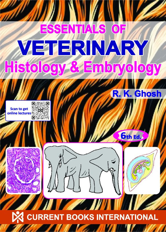


ESSENTIALS of VETERINARY HISTOLOGY & EMBRYOLOGY
-
Rs. 125.00
Rs. 250.00 -
Rs. 600.00
Rs. 800.00 -
Rs. 550.00
-
Rs. 488.00
Rs. 650.00 -
Rs. 450.00
Rs. 600.00 -
Rs. 518.00
Rs. 690.00
Essentials of veterinary histology and embryology
HISTOLOGY
General Histology; Tissues of the Body; Integumentary System; Digestive System; Respiratory System; Urogenital System; Endocrine Glands; Organs of Special Sense
EMBRYOLOGY
General Embryology; Integumentary System; Alimentary System; Respiratory System; Urinary System; Genital System; Endocrine Glands; Organs of Special Senses; Multiple Birth Twinning and Conjoined Twins; Early Development in the Chick Embryo
Reviews & Ratings
Related products
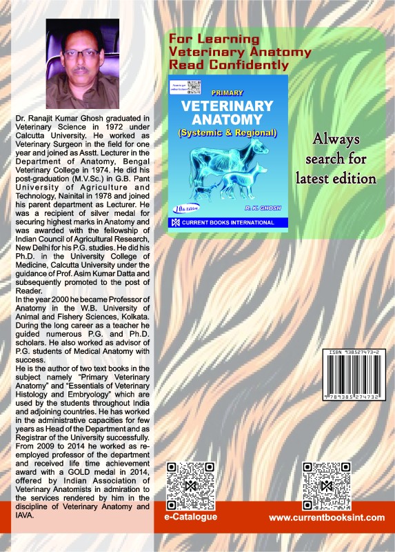
PRIMARY VETERINARY ANATOMY
SURE SUCCESS in VETERINARY PARASITOLOGY
ENCYCLOPEDIC GLIMPSE of VETERINARY PARASITIC DISEASE, 1/ed
-
Rs. 125.00
Rs. 250.00 -
Rs. 600.00
Rs. 800.00 -
Rs. 550.00
-
Rs. 488.00
Rs. 650.00 -
Rs. 450.00
Rs. 600.00 -
Rs. 518.00
Rs. 690.00

